University of Cambridge researchers created four out of the 16 winning images in the recent Wellcome Image Awards 2012.
University of Cambridge researchers created four out of the 16 winning images in the recent Wellcome Image Awards 2012.
The Wellcome Image Awards are unique in that the winners are chosen for their scientific and technical merit as much as for their aesthetic appeal.
Catherine Draycott, Head of Wellcome Images
The Wellcome Image Awards celebrate the best images acquired by the Wellcome Images picture library over the past 18 months. Sixteen winning images were recently selected by a judging panel, based not only on their visual appeal but also their technical excellence and ability to convey the fascination of science.
University of Cambridge researchers were responsible for a quarter of the winning images. Showing bacteria, biofilms, seedlings and chicken embryos, the images created by the researchers display the beauty that can be harnessed using modern technology to magnify the insignificant and undetectable to a microscopic scale, creating an array of surprisingly stunning images.
Now in their 15th year, the Wellcome Image Awards were established to reward contributors to the collection for their outstanding work.
Catherine Draycott, Head of Wellcome Images and a member of the judging panel, said: “The Wellcome Image Awards are unique in that the winners are chosen for their scientific and technical merit as much as for their aesthetic appeal. They offer people a chance to get closer to science and research and see it in a different way, as a source of beauty as well as providing important information about ourselves and the world around us.”
The winning images are now on display at the Wellcome Collection, as well as on the Wellcome Image Awards website.
Displayed below are winning images created by Vincent Pasque, Fernan Federici, Jim Haseloff, Tim Rudge and PJ Steiner.
Credit: Vincent Pasque, University of Cambridge
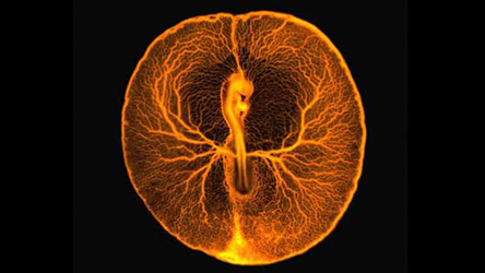
‘Chicken Embryo Vascular System’ by Vincent Pasque
This glowing fluorescence micrograph shows the vascular system of a developing chicken embryo, two days after fertilisation. Injecting fluorescent dextran revealed the entire vasculature used by the embryo to feed itself from the rich underlying yolk inside the egg. At this stage of development, the embryo and its surrounding vasculature are a little smaller than a 5p coin.
Credit: Fernan Federici, Tim Rudge, PJ Steiner and Jim Haseloff
‘Bacteria Biofilm’ by Fernan Federici, Tim Rudge, PJ Steiner and Jim Haseloff
This confocal micrograph, taken as part of a synthetic biology project, shows Bacillus subtilis, a bacterium that is commonly found in soil. Distinct lineages of bacteria expressing different fluorescent proteins were initially mixed randomly on a petri dish. As the bacteria grow, they organise themselves into reproducible patterns and shapes that can be predicted with mathematical models. The abstract organisation of these bacteria create a uniquely artistic image.
Credit: Vincent Pasque
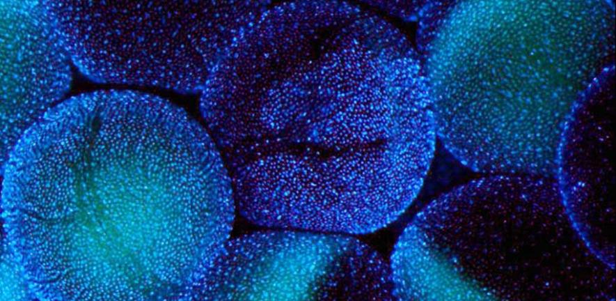
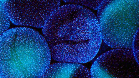
‘African Clawed Frog (Xenopus laevis) Oocytes’ by Vincent Pasque
This beautiful, almost luminous photograph by Vincent Pasque shows stage 5-6 oocytes of an African Clawed Frog, a model organism used in cell and developmental biology research. Each oocyte is surrounded by thousands of follicle cells, shown in the image by staining DNA blue. Tiny blood vessels, which are almost too small to see at this scale, are shown in red. These provide oxygen to the oocyte and follicle cells. The ovary of each adult female African Clawed Frog contains up to 20,000 oocytes, and mature oocytes are approximately 1.2 mm in diameter. Although this sounds miniscule, they are much larger than the eggs of many other species.
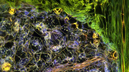
Credit: Fernan Federici and Jim Haseloff
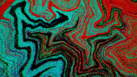
‘Arabidopsis thaliana Seedling’ by Fernan Federici and Jim Haseloff
This photo shows the complex tissue structures within the leaf of an Arabidopsis thaliana seedling. The sample was fixed and stained with propidium iodide, which labels DNA, but was imaged four years later. Over time, oxidation of the stain in different parts of the tissue provides a variation of fluorescent properties that can be excited with distinct wavelengths of light from a confocal microscope. The researchers are using these techniques to investigate cellular architecture in plants and gene activity.

This work is licensed under a Creative Commons Licence. If you use this content on your site please link back to this page.

, similar to the epiblast of the human embryo just after implantation, Credit: University of Cambridge")

