
The first 3D reconstruction of the skull of a 360 million-year-old near-ancestor of land vertebrates has been created by scientists.
The first 3D reconstruction of the skull of a 360 million-year-old near-ancestor of land vertebrates has been created by scientists.
This work is the first stage of a study towards understanding how the earliest tetrapods fed, and that might lead us to what they fed on
Jennifer Clack
A new 3D reconstruction of skull of one of the earliest four-footed vertebrate – which differs from earlier 2D reconstructions – suggests such creatures, which lived their lives primarily in shallow water environments, were more like modern crocodiles than previously thought.
The researchers applied high-resolution X-ray computed tomography (CT) scanning to several specimens of Acanthostega gunnari, one of the ‘four-footed’ vertebrates known as tetrapods which invaded the land during one of the great evolutionary transitions in Earth’s history, 380-360 million years ago. Tetrapods evolved from lobe-finned fishes and display a number of adaptations to help them survive on land.
An iconic fossil species, Acanthostega gunnari is crucial for understanding the anatomy and ecology of the earliest tetrapods. However, after hundreds of millions of years in the ground fossils are often damaged and deformed. No single specimen of Acanthostega preserves a skull that is complete and three-dimensional which has limited scientists’ understanding of how this key animal fed and breathed – until now.
Researchers from Cambridge and Bristol University used specialist software to ‘digitally prepared’ a number of Acanthostega specimens from East Greenland, stripping away layers of rock to reveal the underlying bones.
They uncovered a number of bones deep within the skull, including some that had never before been seen or described, resulting in a detailed anatomical description of the Acanthostega skull.
Once all of the bones and teeth were digitally separated from each other, cracks were repaired and missing elements duplicated. Bones could then be manipulated individually in 3D space. Using information from other specimens, the bones were fitted together like puzzle pieces to produce the first 3D reconstruction of the skull of Acanthostega, with surprising results.
Co-author Dr Laura Porro, formerly of Cambridge’s Department of Zoology and Bristol’s School of Earth Sciences (now at the Royal Veterinary College) said: “Because early tetrapods skulls are often ‘pancaked’ during the fossilization process, these animals are usually reconstructed having very flat heads. Our new reconstruction suggests the skull of Acanthostega was taller and somewhat narrower than previously interpreted, more similar to the skull of a modern crocodile.”
The researchers also found clues to how Acanthostega fed. The size and distribution of its teeth and the shape of contacts between individual bones of the skull (called sutures) suggest Acanthostega may have initially seized prey at the front of its jaws using its large front teeth and hook-shaped lower jaw.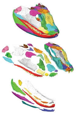
The team say that these new analyses provide fresh clues about the evolution of the jaws and feeding system as the earliest animals with limbs and digits began to conquer the land.
The researchers plan to apply these methods to other flattened fossils of the earliest tetrapods to better understand how these early animals modified their bones and teeth to meet the challenges of living on land.
“This work is the first stage of a study towards understanding how the earliest tetrapods fed, and that might lead us to what they fed on, and give further clues as to when and how they started to feed on land,” said co-author Professor Jennifer Clack from Cambridge’s Zoology Department.
Digital models of the original fossils and the 3D reconstruction are also useful in scientific research and education. They can be accessed by researchers around the world, without risking damage to fragile original fossils and without scientists having to travel thousands of miles to see original specimens. Furthermore, digital models and 3D printouts can be easily and safely handled by students taking courses and by the public during outreach events. The study is published recently in the journal PLOS ONE.
Adapted from a Bristol University press release.
Inset image: 3D model showing the complete skull on top with ‘exploded’ views of the upper and lower jaws below.
The text in this work is licensed under a Creative Commons Licence. If you use this content on your site please link back to this page. For image rights, please see the credits associated with each individual image.


