A new technique that allows embryos to develop in vitro beyond the implantation stage (when the embryo would normally implant into the womb) has been developed by scientists at the University of Cambridge allowing them to analyse for the first time key stages of human embryo development up to 13 days after fertilisation. The technique could open up new avenues of research aimed at helping improve the chances of success of IVF.
A new technique that allows embryos to develop in vitro beyond the implantation stage (when the embryo would normally implant into the womb) has been developed by scientists at the University of Cambridge allowing them to analyse for the first time key stages of human embryo development up to 13 days after fertilisation. The technique could open up new avenues of research aimed at helping improve the chances of success of IVF.
Implantation is a milestone in human development as it is from this stage onwards that the embryo really begins to take shape and the overall body plans are decided
Magdalena Zernicka-Goetz
Once an egg has been fertilised by a sperm, it divides several times to generate a small, free-floating ball of stem cells. Around day three, these stem cells cluster together inside the embryo towards one side; this stage is known as the blastocyst. The blastocyst comprises three cell types: cells that will develop into the future body (which form the ‘epiblast’), cells that will develop into the placenta and allow the embryo to attach to the womb, and cells that form the primitive endoderm that will ensure that the fetus’s organs develop properly and will provide essential nutrients.
This pre-implantation period – so-called as the blastocyst has yet to implant itself into the uterus – has been extensively studied in human embryos using in vitro culture methods. However, on the seventh day of development, the human embryo must implant into the uterus of the mother to survive and to develop further, even though UK law permits embryos to be studied in the laboratory for up to 14 days.
The failure of an embryo to implant is a major cause of early pregnancy loss and yet the cellular and molecular changes that take place in the human embryo at this stage remain unknown. This is because it is impossible to carry out such studies on embryos developing in the womb, and until now there has been no system to culture human embryos in the laboratory beyond day seven.
Today, in parallel papers in Nature and Nature Cell Biology, two international teams report the development of a technique that allows them to culture human embryos outside the body of the mother for an additional six days, up to day 13 of development. This work builds on previous work by Professor Magdalena Zernicka-Goetz’s team from the University of Cambridge on mouse and was funded by the Wellcome Trust.
Using the technique, the researchers have shown that the reorganisation of the embryo that normally takes place during early post-implantation development can be achieved in the lab given the right culture conditions.
Professor Zernicka-Goetz, who led the UK research and is an author on both studies, says: “Implantation is a milestone in human development as it is from this stage onwards that the embryo really begins to take shape and the overall body plans are decided. It is also the stage of pregnancy at which many developmental defects can become acquired. But until now, it has been impossible to study this in human embryos. This new technique provides us with a unique opportunity to get a deeper understanding of our own development during these crucial stages and help us understand what happens, for example, during miscarriage.”
“Embryo development is an extremely complex process and while our system may not be able to fully reproduce every aspect of this process, it has allowed us to reveal a remarkable self-organising capacity of human blastocysts that was previously unknown,” says Dr Marta Shahbazi one of the co-first authors of the study from the University of Cambridge.
The researchers established a system for the in vitro culture of human embryos and, using this technique, followed the development of the embryos up to day 13 of development. Immediately following ‘implantation’, the three cell types that comprise the blastocyst reorganise into a new configuration.
“The stem cells in the epiblast that will form the future body have the remarkable ability to self-organise themselves and create a cavity that represent the basic structure of the early post-implantation human embryo,” says Professor Zernicka-Goetz. “Without this cavity, it would be impossible for the embryo to develop further as it is the basis for its future development. It is also a mechanism that we can study using human embryonic stem cells.”
This cavity was previously thought to arise through a process known as apoptosis, or programmed cell death, but using human embryonic stem cell models, the researchers were able to show that in fact cell death is not required for the cavity formation in human embryos.
“This process is similar to what we have recently observed in mouse embryos, despite the significant differences in the structure of post-implantation embryos in these different mammalian species”, says Professor Zernicka-Goetz. “This suggests it may be a fundamental process conserved across many species.”
Dr Simon Fishel, founder and President of CARE Fertility Group, adds: “This is about much more than just understanding the biology of implantation embryo development. Knowledge of these processes could help improve the chances of success of IVF, of which only around one in four attempts are successful.”
This research has been possible thanks to couples that underwent IVF treatment and decided to donate their surplus embryos to advance our understanding of the early phases of post-implantation human development. The research was licensed by the UK Human Fertilisation and Embryology Authority.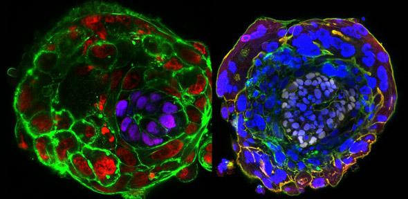
Left image: At day 10 of embryo development the pluripotent stem cells that will generate the future body self-organise to generate a cavity (the pro-amniotic cavity). This configuration is the basis for the subsequent developmental stages and the formation of the body plan. The immunofluorescence image shows a day 10 human embryo cultured in vitro through early post-implantation stages (purple-epiblast, red-nucleus, green-membrane).
Right image: At day 11 of embryo development the pluripotent stem cells that will generate the future body self-organiseto generate a cavity (the pro-amniotic cavity). This configuration is the basis for the subsequent developmental stages and the formation of the body plan. The immunofluorescence image shows a day 11 human embryo cultured in vitro through early post-implantation stages (while-epiblast, blue-nucleus, green-membrane).
Reference
Shahbazi, MN et al. Self-organisation of the human embryo in the absence of maternal tissues. Nature Cell Biology; 4 May 2016; DOI: 10.1038/ncb3347
Deglincerti, A et al. Self-organization of the in vitro attached human embryo. Nature; 4 May 2016

The text in this work is licensed under a Creative Commons Attribution 4.0 International License. For image use please see separate credits above.


")
. Trophoblast cells are invading out of the organoid, mimicking placental cells invading the uterus in the early weeks of pregnancy., Credit: Friedrich Miescher Institute/University of Cambridge")