New study examines thousands of brains from two decades of research to reveal differences between male and female brain structure.
New study examines thousands of brains from two decades of research to reveal differences between male and female brain structure.
For the first time we can look across the vast literature and confirm that brain size and structure are different in males and females
Amber Ruigrok
Reviewing over 20 years of neuroscience research into sex differences in brain structure, a Cambridge University team has conducted the first meta-analysis of the evidence, published this week in the journal Neuroscience and Biobehavioral Reviews.
The team, led by doctoral candidate Amber Ruigrok and Professors John Suckling and Simon Baron-Cohen in the Department of Psychiatry, performed a quantitative review of the brain imaging literature testing overall sex differences in total and regional brain volumes. They searched all articles published between 1990 and 2013. A total of 126 articles were included in the study, covering brains from individuals as young as birth to 80 years old.
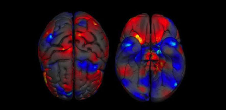
They found that males on average have larger total brain volumes than women (by 8-13%). On average, males had larger absolute volumes than females in the intracranial space (12%; >14,000 brains), total brain (11%; 2,523 brains), cerebrum (10%; 1,851 brains), grey matter (9%; 7,934 brains), white matter (13%; 7,515 brains), regions filled with cerebrospinal fluid (11.5%; 4,484 brains), and cerebellum (9%; 1,842 brains). Looking more closely, differences in volume between the sexes were located in several regions. These included parts of the limbic system, and the language system.
Specifically, males on average had larger volumes and higher tissue densities in the left amygdala, hippocampus, insular cortex, putamen; higher densities in the right VI lobe of the cerebellum and in the left claustrum; and larger volumes in the bilateral anterior parahippocampal gyri, posterior cingulate gyri, precuneus, temporal poles, and cerebellum, areas in the left posterior and anterior cingulate gyri, and in the right amygdala, hippocampus, and putamen.
By contrast, females on average had higher density in the left frontal pole, and larger volumes in the right frontal pole, inferior and middle frontal gyri, pars triangularis, planum temporale/parietal operculum, anterior cingulate gyrus, insular cortex, and Heschl’s gyrus; bilateral thalami and precuneus; the left parahippocampal gyrus, and lateral occipital cortex.
The results highlight an asymmetric effect of sex on the developing brain. Amber Ruigrok, who carried out the study as part of her PhD, said: “For the first time we can look across the vast literature and confirm that brain size and structure are different in males and females. We should no longer ignore sex in neuroscience research, especially when investigating psychiatric conditions that are more prevalent in either males or females.”
Professor Suckling added: “The sex differences in the limbic system include areas often implicated in psychiatric conditions with biased sex ratios such as autism, schizophrenia, and depression. This new study may therefore help us understand not just typical sex differences but also sex-linked psychiatric conditions. It is important to note that we only investigated sex differences in brain structure, so we cannot infer anything about how this relates to behaviour or brain function. Integrating across different levels will be an important goal for future research.”
Professor Baron-Cohen commented: “Although these very clear sex differences in brain structure may reflect an environmental or social factor, from other studies we know that biological influences are also important, including prenatal sex steroid hormones (such as foetal testosterone) as well as sex chromosome effects. Such influences need to be teased out, one by one.”
Dr Meng-Chuan Lai, another member of the team, noted: “The advantage of conducting a meta-analysis is that we can summarise the best knowledge from a vast, heterogeneous literature, with a very large sample size. However, we found a bias in the existing literature towards the use of volunteers over 18 years old, probably because this is the easiest age group to recruit and to brain scan. We need more research exploring brain development over the entire lifespan, especially in the early, formative years”.
This study was supported by the Medical Research Council, the Wellcome Trust, the Autism Research Trust, the Dr Hendrik Müller Vaderlandsch Fonds, the Carolus Magnus Fonds under the Prins Bernard Cultuurfonds, and the Waterloo Foundation. It was conducted in association with the NIHR CLAHRC for Cambridgeshire and Peterborough NHS Foundation Trust.
Professor Simon Baron-Cohen will be speaking on ‘Do hormones in the womb affect how your brain and mind develop’ on Saturday 15 March at the Cambridge Science Festival. For more information about the Festival and to book tickets for Professor Baron-Cohen’s talk, please visit: www.cam.ac.uk/science-festival

This work is licensed under a Creative Commons Licence. If you use this content on your site please link back to this page.


