‘Synthetic’ embryo with brain and beating heart grown from stem cells by Cambridge scientists

Researchers from the University of Cambridge have created model embryos from mouse stem cells that form a brain, a beating heart, and the foundations of all the other organs of the body – a new avenue for recreating the first stages of life.
The team, led by Professor Magdalena Zernicka-Goetz, developed the embryo model without eggs or sperm, and instead used stem cells – the body’s master cells, which can develop into almost any cell type in the body.
The researchers mimicked natural processes in the lab by guiding the three types of stem cells found in early mammalian development to the point where they start interacting. By inducing the expression of a particular set of genes and establishing a unique environment for their interactions, the researchers were able to get the stem cells to ‘talk’ to each other.
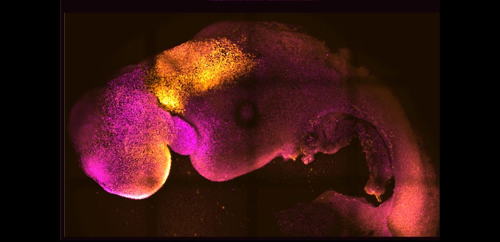
The stem cells self-organised into structures that progressed through the successive developmental stages until they had beating hearts and the foundations of the brain, as well as the yolk sac where the embryo develops and gets nutrients from in its first weeks. Unlike other synthetic embryos, the Cambridge-developed models reached the point where the entire brain, including the anterior portion, began to develop.
This is a further point in development than has been achieved in any other stem cell-derived model.


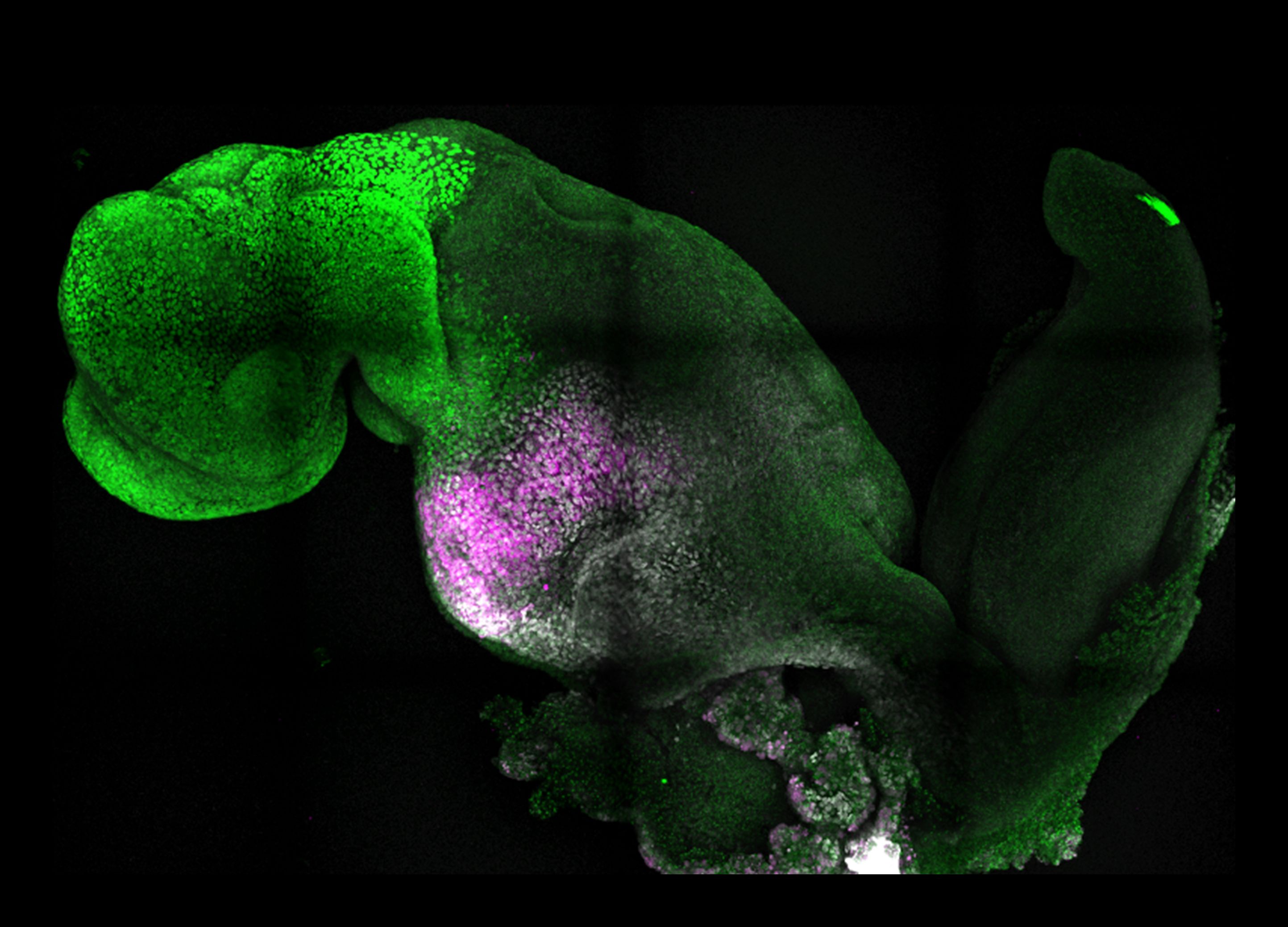
Natural (top) and synthetic (bottom) embryos side by side to show comparable brain and heart formation. Image credit: Amadei and Handford
Natural (top) and synthetic (bottom) embryos side by side to show comparable brain and heart formation. Image credit: Amadei and Handford
The team say their results, the result of more than a decade of research that progressively led to more and more complex embryo-like structures and reported today in the journal Nature, could help researchers understand why some embryos fail while others go on to develop into a healthy pregnancy. Additionally, the results could be used to guide repair and development of synthetic human organs for transplantation.
“Our mouse embryo model not only develops a brain, but also a beating heart, all the components that go on to make up the body,” said Zernicka-Goetz, Professor in Mammalian Development and Stem Cell Biology in Cambridge’s Department of Physiology, Development and Neuroscience, adding:
“It’s just unbelievable that we’ve got this far. This has been the dream of our community for years, and a major focus of our work for a decade, and finally we’ve done it.”
For a human embryo to develop successfully, there needs to be a ‘dialogue’ between the tissues that will become the embryo, and the tissues that will connect the embryo to the mother. In the first week after fertilisation, three types of stem cells develop: one will eventually become the tissues of the body, and the other two support the embryo’s development. One of these extraembryonic stem cell types will become the placenta, which connects the foetus to the mother and provides oxygen and nutrients; and the second is the yolk sac, where the embryo grows and where it gets its nutrients from in early development.
Many pregnancies fail at the point when the three types of stem cells begin to send mechanical and chemical signals to each other, which tell the embryo how to develop properly.
“So many pregnancies fail around this time, before most women realise they are pregnant,” said Zernicka-Goetz, who is also Professor of Biology and Biological Engineering at Caltech. “This period is the foundation for everything else that follows in pregnancy. If it goes wrong, the pregnancy will fail.”
Over the past decade, Professor Zernicka-Goetz’s group in Cambridge has been studying these earliest stages of pregnancy, in order to understand why some pregnancies fail and some succeed.
“The stem cell embryo model is important because it gives us accessibility to the developing structure at a stage that is normally hidden from us due to the implantation of the tiny embryo into the mother’s womb."
Zernicka-Goetz added: “This accessibility allows us to manipulate genes to understand their developmental roles in a model experimental system.”
To guide the development of their synthetic embryo, the researchers put together cultured stem cells representing each of the three types of tissue in the right proportions and environment to promote their growth and communication with each other, eventually self-assembling into an embryo.
The researchers found that the extraembryonic cells signal to embryonic cells by chemical signals but also mechanistically, or through touch, guiding the embryo’s development.
“This period of human life is so mysterious, so to be able to see how it happens in a dish – to have access to these individual stem cells, to understand why so many pregnancies fail and how we might be able to prevent that from happening – is quite special,” said Zernicka-Goetz. “We looked at the dialogue that has to happen between the different types of stem cell at that time – we’ve shown how it occurs and how it can go wrong.”
A major advance in the study is the ability to generate the entire brain, in particular the anterior part, which has been a major goal in the development of synthetic embryos.
This works in Zernicka-Goetz’s system because this part of the brain requires signals from one of the extraembryonic tissues to be able to develop. The team thought that this might be taking place from their 2018 and 2021 studies, which used the same component cells to develop into embryos at a slightly earlier stage. Now, by pushing development just one day further, they can definitively say that their model is the very first to signal development of the anterior, and in fact the whole, brain.
“This opens new possibilities to study the mechanisms of neurodevelopment in an experimental model,” said Zernicka-Goetz. “In fact, we demonstrate the proof of this principle in the paper by knocking out a gene already known to be essential for formation of the neural tube, precursor of the nervous system, and for brain and eye development. In the absence of this gene, the synthetic embryos show exactly the known defects in brain development as in an animal carrying this mutation. This means we can begin to apply this kind of approach to the many genes with unknown function in brain development.”
While the current research was carried out in mouse models, the researchers are developing similar human models with the potential to be directed towards the generation of specific organ types - to understand mechanisms behind crucial processes that would be otherwise impossible to study in real embryos. At present, UK law permits human embryos to be studied in the laboratory only up to the 14th day of development.
If the methods developed by Zernicka-Goetz’s team are shown to be successful with human stem cells in future, they could also be used to guide development of synthetic organs for patients awaiting transplants.
“There are so many people around the world who wait for years for organ transplants,” said Zernicka-Goetz. “What makes our work so exciting is that the knowledge coming out of it could be used to grow correct synthetic human organs to save lives that are currently lost. It should also be possible to affect and heal adult organs by using the knowledge we have on how they are made.
Professor Magdalena Zernicka-Goetz has made an incredible scientific breakthrough.
The creation of synthetic mouse embryos in a test tube that develop brains and beating hearts, starting only with embryonic stem cells, is the culmination of a decade’s work.
Magda explains:

I’m fascinated by the mystery of how embryos work. Every embryo follows a similar journey: one cell becomes many, then they communicate with each other and arrange themselves to form a structure that will provide a blueprint for all adult body parts. But how do embryo cells decide their fate, how do they know where to go and what to do? How do they form the right parts in the right place at the right time?


Image credit: Science Photo Library
Image credit: Science Photo Library
Building the first ‘synthetic embryo’ models was a process we achieved step by step. Setting out, we knew that embryonic stem cells could be cultured indefinitely in the lab, and that when they’re injected into an embryo they can potentially contribute to any tissue in the adult organism. The challenge was to guide them to develop into a complete embryo. In addition to the embryonic stem cells we used two kinds of extraembryonic tissue: one of which forms the placenta and the other a sac in which the embryo develops. These tissues are very important, as they send signals to the embryo to develop all its parts at the right time and in the right place.
Combining stem cells representing each of these three types of tissue is easier said than done. We had to find an environment where all three distinct cell types could grow and communicate with each other. And we had to find the right proportions of each cell type, and add them in the right sequence. Once we established these basic principles, the stem cells did the rest: they self-organised to progress through successive developmental stages until they had beating hearts and the foundations for a brain.
The key to our achievement was thinking outside the conventional box. The majority of embryo model studies focus on embryonic stem cells, but don’t consider the significant role of extraembryonic cells. We mixed the right proportions of both embryonic and extraembryonic stem cells. Extraembryonic cells signal to embryonic cells through different means, by chemical signals but also mechanistically ‘by touch’. Our studies are helping to understand these signalling events.

Image credit: Simon Zernicki-Glover
Image credit: Simon Zernicki-Glover
We are developing an analogous model of the human embryo, to understand the mechanisms behind crucial processes that would be otherwise impossible to study. This is important because the great majority of human pregnancies fail at this developmental stage, due to causes that we don’t understand. It will also allow us to identify factors permitting development of healthy human tissues as they form different organs.
Creating the new ‘synthetic embryo’ has taught us a lot about the mechanisms by which the embryo builds itself. We learnt how the extraembryonic tissues direct the embryonic stem cells along the right pathways to signal formation of the correct structures; how cells move between compartments as the multi-layered body plan arises; and how this correctly sets the scene for neurulation - the process where tissue folds to form the neural tube and, in turn, the brain and spinal cord.
This model gives us access to the developing structure at a stage that’s normally hidden from us, when the tiny embryo implants into the mother’s womb. Our model does not have to implant to develop, so it remains completely visible to us, allowing us to see the embryo’s progression through that developmental stage. This accessibility allows us to manipulate genes to understand their developmental roles in a model experimental system.


Natural (top) and synthetic (bottom) embryos side by side to show comparable brain and heart formation. Image credit: Amadei and Handford
Natural (top) and synthetic (bottom) embryos side by side to show comparable brain and heart formation. Image credit: Amadei and Handford
It’s certainly true that carrying out this type of work requires passion and resilience. I grew up in Poland under a Communist regime, which meant that traveling wasn’t allowed and thinking differently was not encouraged. There was immense social pressure to conform, and a lot of us rebelled against that. A silver lining of this was a desire to think independently and to persevere despite discouragement. That shaped me as a scientist too.
When I started my research group in Cambridge, I established ways to study the ‘developmental black box’ - the development of the embryo at the time of implantation. My mentors had discouraged me from pursuing it during my PhD because they were concerned it would be difficult to shine light inside this ‘box.’ But I was so taken by the question of how the embryo self-organises that I didn’t give up and, inch by inch, we have worked our way forward.
My advice to all young scientists is to follow your heart. Study a topic that inspires you, and choose an advisor who can be supportive of your style of work. In my opinion, it’s important to guide young scientists in the lab, but also to give them space to explore their individuality. My experience is that the challenges for female scientists increase as they progress through their careers. At later stages, it’s critical to have mentors who understand not only science but also how to balance it with everyday life, including starting a family.


Image credit: Science Photo Library
Image credit: Science Photo Library
During my own pregnancy, I was shocked when an early screening showed abnormalities. The sampling was of extraembryonic cells so I waited for the amniocentesis, which samples foetal cells that have fallen into the amniotic fluid. These were normal, which put my mind at ease. The experience led me to study mosaic aneuploidy - a condition in which the embryo has cells with the wrong number of chromosomes alongside chromosomally normal cells. Incredibly, we found that these abnormal cells can be eliminated, and the normal, healthy cells compensate for their absence. For some reason this mechanism doesn’t operate in the tissues that build the placenta, and we’re still trying to understand why and how.
Science is demanding, it’s hard work and it takes away most of your waking hours. I switch off by watching films - I watch a lot foreign films in Polish, French, and Danish, and documentaries and art films. But when I want to lose myself in another narrative, I watch dramas. I’m also a recent convert to gardening, where I can encourage the successful development of other life forms!
It is an incredible feeling and a privilege to have this direct insight into the origins of a new life. It’s like discovering a new planet that we didn't know existed.
Magdalena Zernicka-Goetz is Professor of Mammalian Development and Stem Cell Biology at the University of Cambridge and Bren Professor of Biology and Biological Engineering at the California Institute of Technology.
In 2020 she published ‘The Dance of Life: Symmetry, Cells and How We Become Human,’ with Penguin.


The research was supported in part by the European Research Council and Wellcome and NIH Pioneer Awards to Magdalena Zernicka-Goetz. Zernicka-Goetz is a Fellow of Sidney Sussex College, Cambridge.
Reference
Gianluca Amadei et al. ‘Synthetic embryos complete gastrulation to neurulation and organogenesis.’ Nature (August 2022). DOI: 10.1038/s41586-022-05246-3
The text in this work is licensed under a Creative Commons Attribution 4.0 International License
