
Putting plants
under the
microscope


Humans have been making use of plants for thousands of years. Plants make an enormous range of chemicals, often in large amounts - and use these to make complex structures and materials. Despite major advances in synthetic biochemistry, many important medicines and other valuable compounds such as oils, dyes, flavours and fragrances can still only be sourced from plants.
But we’ve barely scratched the surface of their potential as green factories for new biochemicals and biomaterials.
The University of Cambridge is home to one of the largest concentrations of plant scientists anywhere in the world. And as the manager overseeing the largest collection of advanced microscopes in Cambridge, Dr Raymond Wightman at the Sainsbury Laboratory (SLCU) has a front row seat. These machines are helping researchers discover the most incredible details about plants’ structure and function.

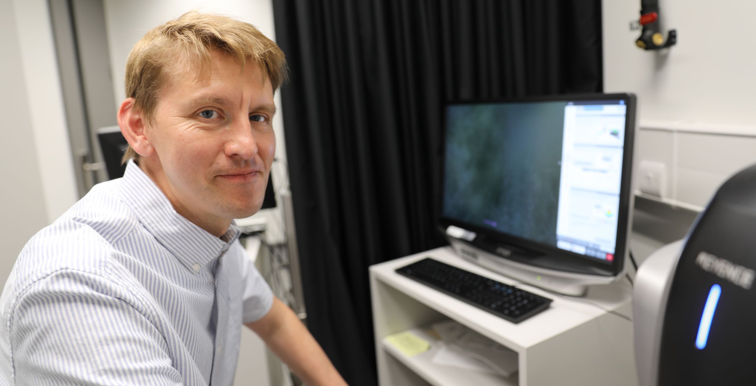
Raymond Wightman at the Sainsbury Lab
Raymond Wightman at the Sainsbury Lab
They include confocal and fluorescence microscopes, equipment for digital imaging, light-sheet, cryo-scanning electron microscopy (cryo-SEM), atomic force microscopy and chemical imaging.
Each microscope has its specific use. For example, the cryo-SEM’s high magnification enabled plant cell wall pectin nanofilaments, which are 1,000 times thinner than a human hair, to be seen for the first time. While the SLCU research team of Alexander Jones is developing biosensors for confocal microscopes that enable plant scientists to track in real time the molecules behind plant growth.
Since the Sainsbury Laboratory opened 10 years ago at the University of Cambridge, its microscopy facility has underpinned many of the Lab’s major discoveries.
The facility supports over 80 plant scientists at the Sainsbury Lab who study the mechanisms underlying plant growth and development, as well as scientists from across the University working on plant-related research.
Each year, Cambridge plant scientists rack up more than 12,000 hours on the Lab’s 40 microscopes, including 15 advanced microscopes.
With support from Wightman and senior imaging technician Gareth Evans, researchers have used the microscopes to track living cells as they divide, grow and change shape to make new flowers. They have observed the activation (and de-activation) of important regulatory genes like those that protect plants from pathogens, and measured the mechanical forces that influence cell shape and tissue organisation. They have also identified petal surface structures that aid pollination, and visualised the outputs of plant hormones within intact seedlings.

Live tracking of a growing root, using confocal microscope and ABACUS biosensor developed at SLCU, shows hormone accumulation in Arabdiopsis roots exposed to the plant hormone abscisic acid. Credit Jim Rowe.
Live tracking of a growing root, using confocal microscope and ABACUS biosensor developed at SLCU, shows hormone accumulation in Arabdiopsis roots exposed to the plant hormone abscisic acid. Credit Jim Rowe.
And it is not only the plant scientists who benefit. Thanks to Wightman’s encouragement, they have been joining forces with horticulturists, chemists, biochemists and material scientists to delve into the world of plant biochemistry and biomaterials.
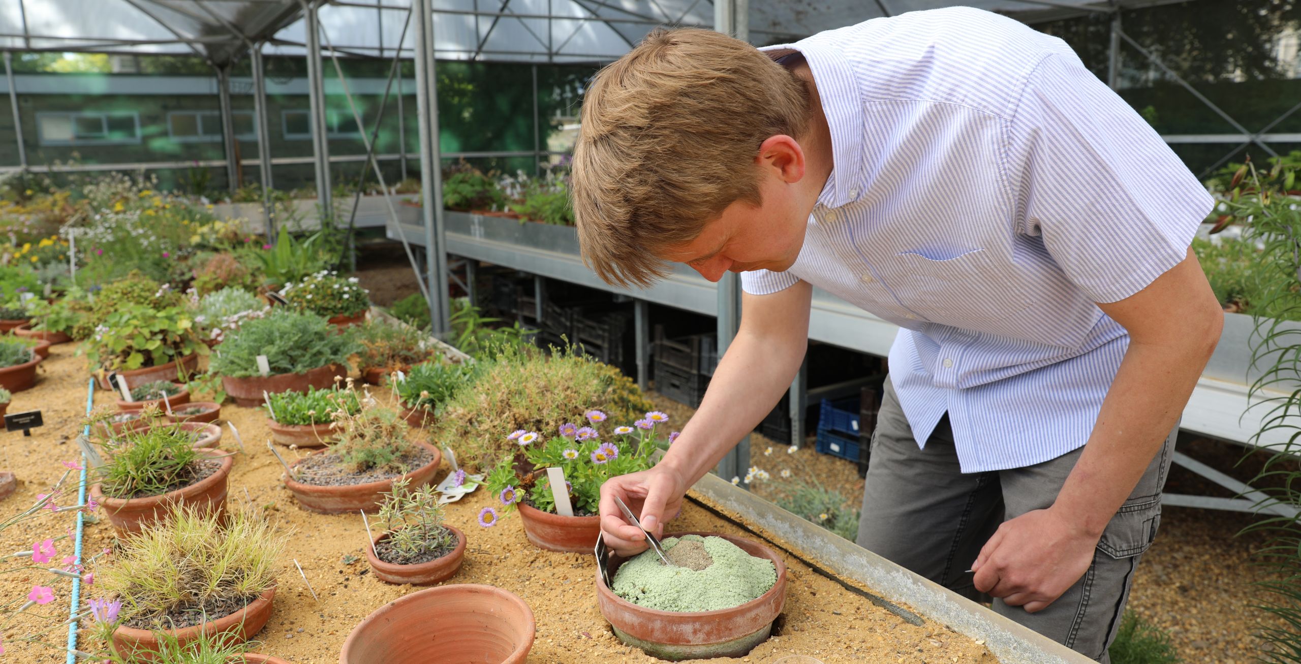
The venture into exploring biomaterials began when Wightman approached the Cambridge University Botanic Garden (CUBG) five years ago to request thick leaf samples to test a new upgrade to one of the microscopes. These samples would not normally be amenable to microscopy.
What started as a casual request has turned into a fruitful ongoing collaboration that is uncovering potential new materials for industrial and medical applications.
Living bio-factories
The Sainsbury Laboratory is located within the 16-hectare grounds of the Botanic Garden in central Cambridge, home to a living collection of 8,000 cultivated plant species - including 14,000 individual accessions, which are separate samples of the same species collected from different parts of the world to capture the species’ diversity.
While tropical rainforests are often talked about as being rich sources of undiscovered pharmaceuticals, alpine plants growing in the harsh mountainous regions of the world could also be a source of potential new medicines and biomaterials.
The Botanic Garden has more than 2,000 species from the alpine and rocky regions of the world in its collection - and this is where Wightman's collaboration began. His focus was on specimens with unusual characteristics that had not received much past scientific attention.
Looking at these alpine plants with advanced imaging and chemical analysis equipment is turning up previously unseen microscopic structures and unusual chemical compounds.
“Alpine plants grow in very harsh conditions at high altitudes above where trees can survive. They can withstand below freezing temperatures, high winds, low humidity and high UV,” says Paul Aston, the Botanic Garden Alpine and Woodland Supervisor.
“There are many things plants make that we don’t yet know about, and this is especially true for alpine plants – which have many unusual adaptations to the harsh environments they live in.”
Manufacturing rare minerals
The first set of alpine plants to be studied were from the CUBG’s national collection of European Saxifraga species, where the researchers found a rare and useful mineral called vaterite coating their leaves.
Researchers talk about their discovery of vaterite coating the leaves of Saxifraga
“Vaterite is an unstable form of calcium carbonate, and in high humidity it usually transforms into more common forms of calcium carbonate like calcite,” says Wightman. “As such, vaterite is rarely found on Earth and is more likely to be found in outer-space on meteorites. Small amounts of vaterite crystals have been found in some sea and freshwater crustaceans, bird eggs and the inner ears of salmon. And now we’ve found it in large concentrations on these alpine plants.”
This discovery is of interest to pharmaceutical and manufacturing industries as vaterite has a number of applications in medicine and printing, but is challenging to manufacture. Saxifraga plants might hold some secrets on how to manufacture vaterite and maintain its stability.y.
Bio-inspired optics
Microscopy examination of Saxifraga plants also turned up some novel cell structures. “Saxifraga scardica has a special tissue surrounding the leaf edge that appears to deflect light from the edge into the leaf,” says Wightman. “The cells appear to be producing novel cell wall structures to achieve this deflection. This may be to help the plant collect more light, particularly if it is growing in partly shaded environments.”

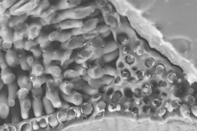
Novel cell wall structures in Saxifraga
Novel cell wall structures in Saxifraga
The team believes the novel cell wall structures of Saxifrages could also one day help inform the manufacture of new bio-inspired optical devices and photonic structures for industry, such as communication cables and fibre optics.

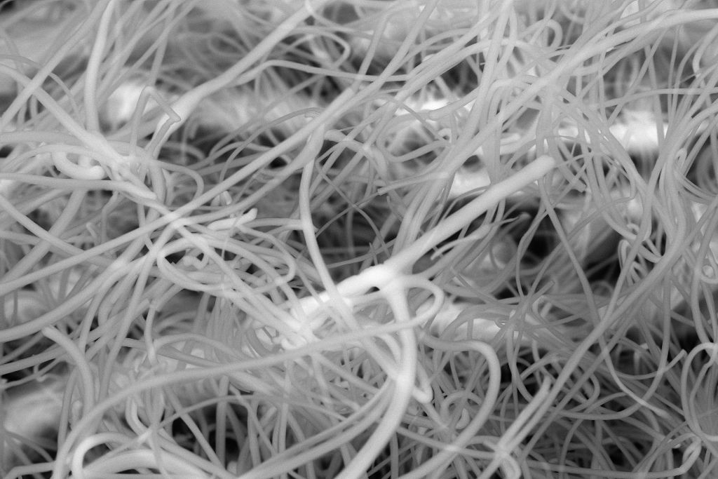
Plants that spin their own wool
Another alpine species from the CUBG collection, Dionysia tapetodes, has been found to spin its own woolly flavonoid covering. Native to central Asia in the mountainous regions around eastern Iran, Dionysia tapetodes is in the primula family. The small perennial cushion-shaped plant has bright yellow flowers and its leaves are covered in long silky fibres that resemble fine cobwebs called woolly farina.
Learn more about the alpine plant Dionysia tapetodes that spins its own 'wool', which covers its leaves. Opening image credit: Matthieu Bourdon
In collaboration with colleagues from the Department of Chemistry and the Cambridge Advanced Imaging Centre, SLCU researchers discovered that flavonoid wool was being ejected via holes in specialised glandular cells on leaves.

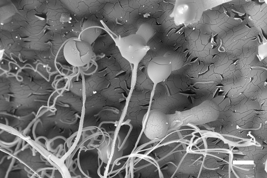
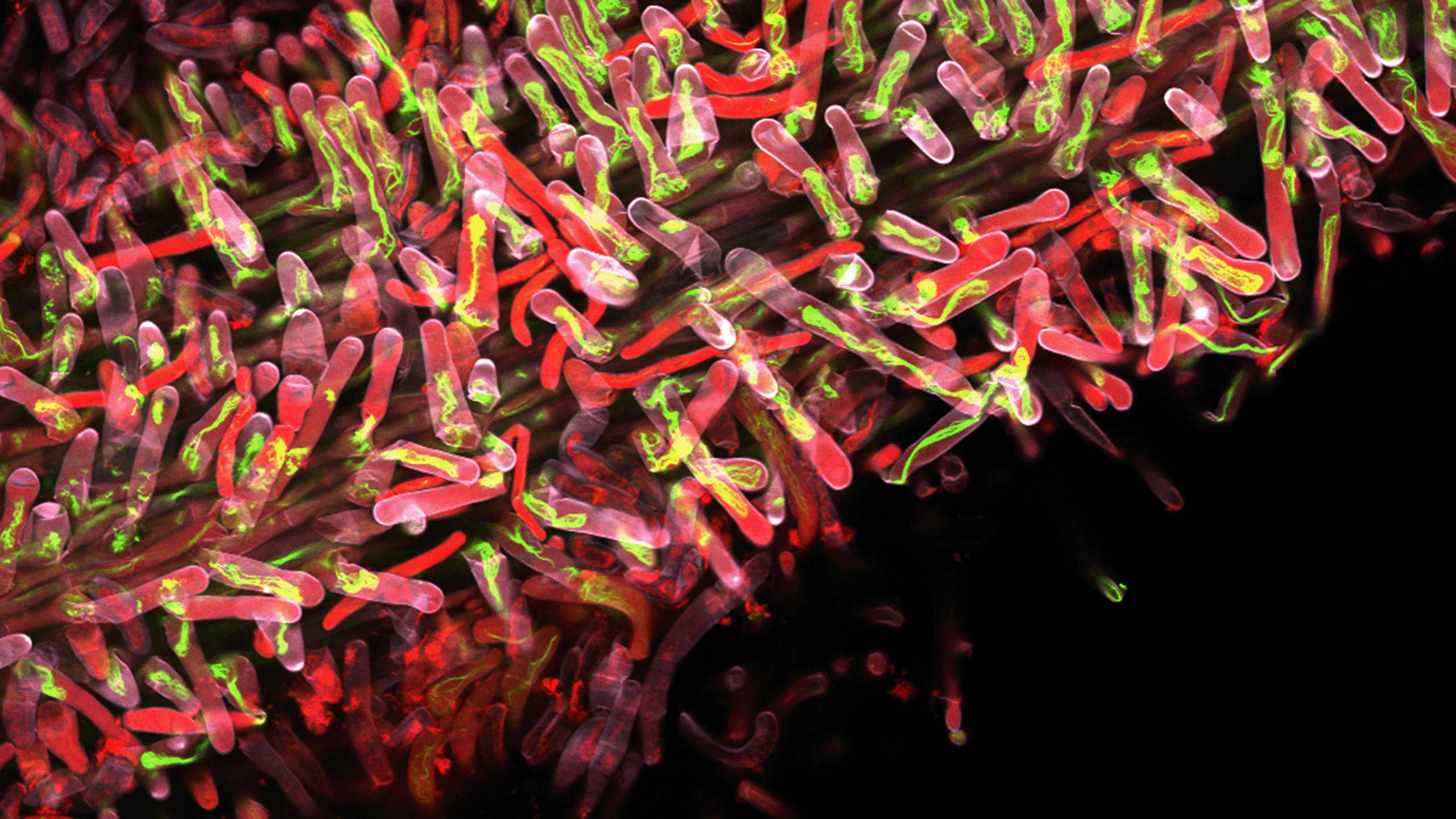
Scanning electron microscope image of Dionysia tapetodes, showing woolly fibres emerging. Credit: Raymond Wightman.
Scanning electron microscope image of Dionysia tapetodes, showing woolly fibres emerging. Credit: Raymond Wightman.
Flavonoids are used widely in food and recognised for their anti-inflammatory and antioxidant properties. The team is interested in further exploring the properties of these flavone fibres to determine if they might be a useful biomaterial.

Growing buildings
In addition to novel biochemicals, plants have been supplying us with construction materials for millennia. Architects and engineers are increasingly considering wood as a lighter and more sustainable construction alternative to steel and concrete.
This has led to a renewed interest in understanding the basic structure and mechanical properties of wood, and how to engineer wood for use in major superstructures like skyscrapers.
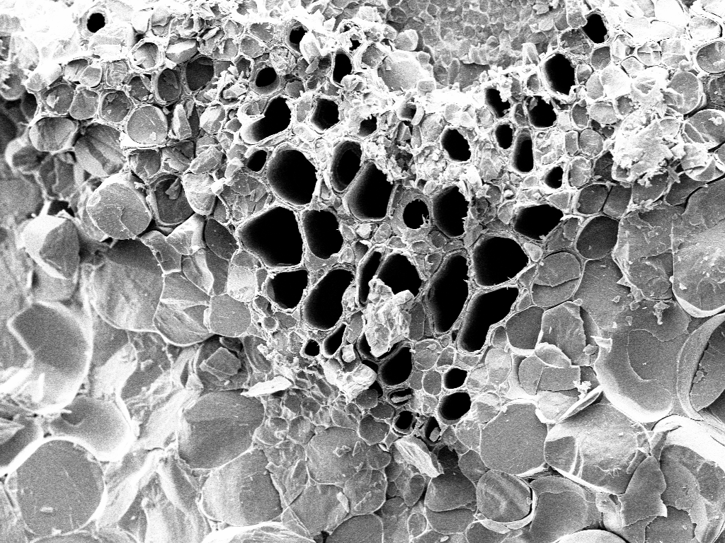
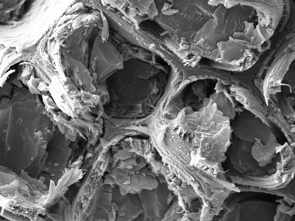
Professor Paul Dupree’s group at Cambridge’s Department of Biochemistry study the properties of plant cell walls and are involved in the Natural Material Innovation Centre where biochemists, plant scientists, architects, mathematicians and chemists from the University of Cambridge are working towards better understanding of wood structure, modification and application.
Using wood samples from three trees – spruce, ginkgo and poplar – from the Botanic Garden, the SLCU and Biochemistry team examined the molecular architecture of wood using snap-frozen samples under a scanning electron microscope (cryo-SEM). The imaging of the inner structure of cell walls helped reveal information about structures, called macrofibrils, and how they contribute to cell wall strength.
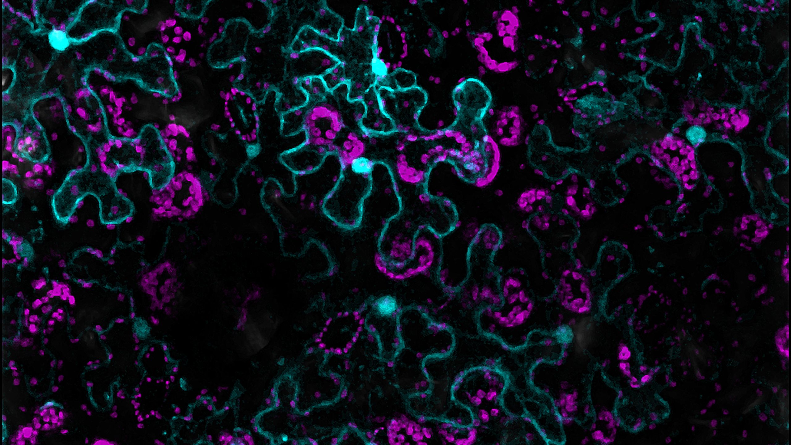
Zooming in even closer to the cell wall structure, the SLCU Microscopy Facility collaborated with plant scientists in Paris, discovering new filamentous structures within plant cell walls that help plants build their complex shapes.

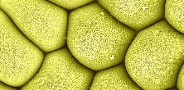
Researchers used sound to analyse the wavy puzzle-shaped cells on a plant’s epidermis. Credit: Raymond Wightman & Alexis Peaucelle.
Researchers used sound to analyse the wavy puzzle-shaped cells on a plant’s epidermis. Credit: Raymond Wightman & Alexis Peaucelle.
“Combining two types of high-performance microscopes, we identified pectin nanofilaments aligned in columns along the edge of the cell walls of plants,” said Wightman. “The filaments, which are 1,000 times thinner than a human hair, had only ever been synthesised in a lab, but never observed in nature until now.”
Not only did they discover these new structures, they also demonstrated, through computational modelling, that the structures could drive cell shape – and even cell growth – independent of pressure within the cell.

Wightman envisages further discoveries in plant and human health will come with further improvements in microscope technology, and is looking forward to further exploring the incredibly diverse living collection in the Cambridge University Botanic Garden.
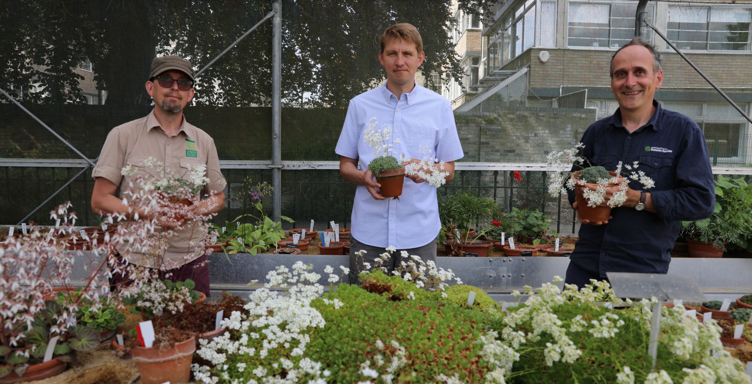
Ray Wightman (centre) with colleagues Simon Wallis (left) and Paul Aston (right), holding Saxifrage from the Botanic Garden's alpine collection
Ray Wightman (centre) with colleagues Simon Wallis (left) and Paul Aston (right), holding Saxifrage from the Botanic Garden's alpine collection

More information at: www.slcu.cam.ac.uk/facilities/microscopy
Plant and microscopy images used here with thanks to the scientists at the Sainsbury Laboratory.
Images of Raymond Wightman and colleagues by Lloyd Mann.
