Human embryo-like model created from human
stem cells


Scientists from the University of Cambridge, in collaboration with the Hubrecht Institute in The Netherlands, have developed a new model to study an early stage of human development using human embryonic stem cells.
The model resembles some key elements of an embryo at around 18-21 days old and allows the researchers to observe the processes underlying the formation of the human body plan never directly observed before. Understanding these processes holds potential to reveal the causes of human birth defects and diseases, and to develop tests for these in pregnant women.
The body plan, or blueprint of an organism, arises through a process called ‘gastrulation’. During gastrulation, three distinct layers of cells are formed in the embryo that will later give rise to all the body’s major systems: the ectoderm will make the nervous system, mesoderm the muscles, and endoderm the gut.
Gastrulation is often referred to as the ‘black box’ period of human development, because legal restrictions prevent the culture of human embryos in the lab beyond day 14, when the process starts. This limit was set to fall at the stage where the embryo can no longer form a twin.
Many birth defects originate during this ‘black box’ period, with causes including alcohol, medications, chemicals and infections. A better understanding of human gastrulation could also shed light on many medical issues including infertility, miscarriage, and genetic disorders.
“Our model produces part of the blueprint of a human. It’s exciting to witness the developmental processes that until now have been hidden from view - and from study.”
Published today in the journal Nature, the report describes a method of using human embryonic stem cells to generate a three-dimensional assembly of cells, called gastruloids, which differentiate into three layers organised in a manner that resembles the early human body plan.
To make gastruloids in the lab, defined numbers of human embryonic stem cells were placed in small wells, where they formed tight aggregates. After treatment with chemical signals, the gastruloids were seen to lengthen along a head to tail axis, known as the anteroposterior axis, turning on genes in specific patterns along this axis that reflect elements of a mammalian body plan.

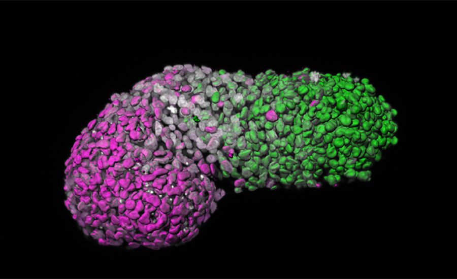
Image analysis of human gastruloid showing ‘anteroposterior’ patterning. Green is the posterior part, similar to the tail-end of an embryo; magenta is the anterior part, similar to developing heart cells; grey marks DNA.
Image analysis of human gastruloid showing ‘anteroposterior’ patterning. Green is the posterior part, similar to the tail-end of an embryo; magenta is the anterior part, similar to developing heart cells; grey marks DNA.
Model organisms including mice and zebrafish have previously enabled scientists to gain some insights into human gastrulation. However, these models may behave differently to human embryos when the cells start to differentiate. Animal models can respond differently to certain drugs: the anti-morning sickness drug thalidomide, for example, passed clinical trials after testing in mice but subsequently led to severe birth defects in humans. For this reason it is important to develop better models of human development.
Gastruloids do not have the potential to develop into a fully-formed embryo. They do not have brain cells or any of the tissues needed for implantation in the womb. This means they would never be able to progress past the very early stages of development, and therefore conform to current ethical standards.

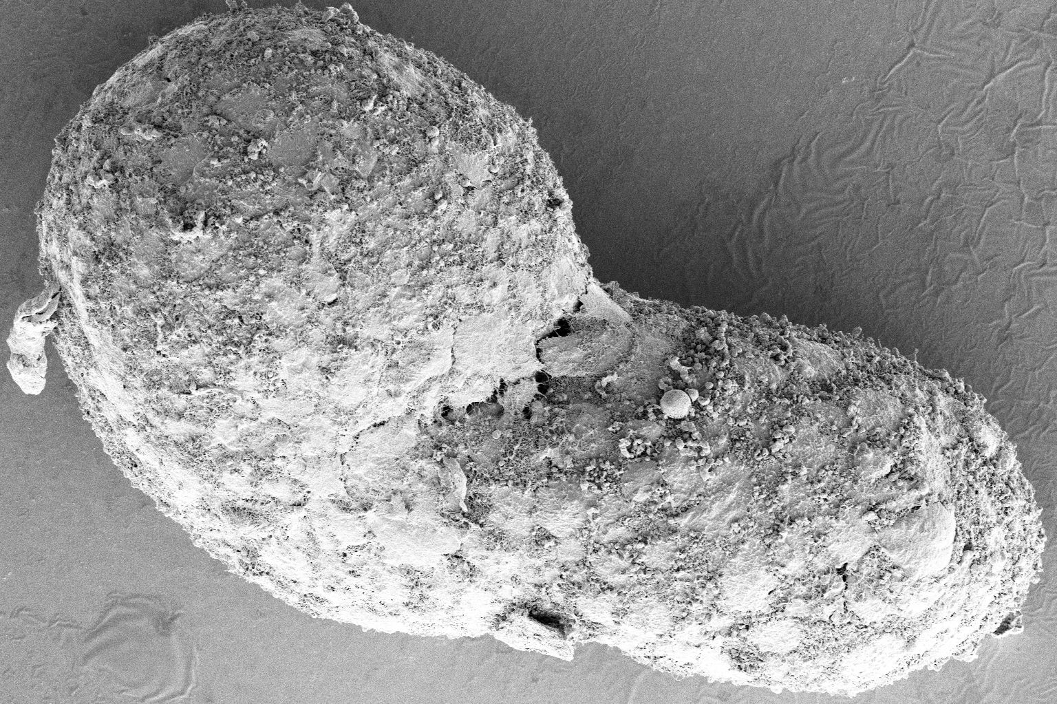
72 hour human gastruloid at high magnification, imaged using Scanning Electron Microscopy (SEM).
72 hour human gastruloid at high magnification, imaged using Scanning Electron Microscopy (SEM).
By looking at which genes were expressed in these human gastruloids at 72 hours of development, the researchers found a clear signature of the event that gives rise to important body structures such as thoracic muscles, bone and cartilage, but they do not develop brain cells.
The researchers judged the equivalent human embryonic age of the gastruloids by comparing them to the Carnegie Collection of Embryology. This official collection contains a continuum of human embryos, including day-by-day growth over the first eight weeks. They suggest that gastruloids partially resemble 18-21 day old human embryos.
“This is a hugely exciting new model system, which will allow us to reveal and probe the processes of early human embryonic development in the lab for the first time.”
Moris added: “Our system is a first step towards modelling the emergence of the human body plan, and could prove useful for studying what happens when things go wrong, such as in birth defects.”
The new ‘gastruloid’ model, created from human stem cells, holds huge potential to understand causes of birth defects and improve disease modelling.

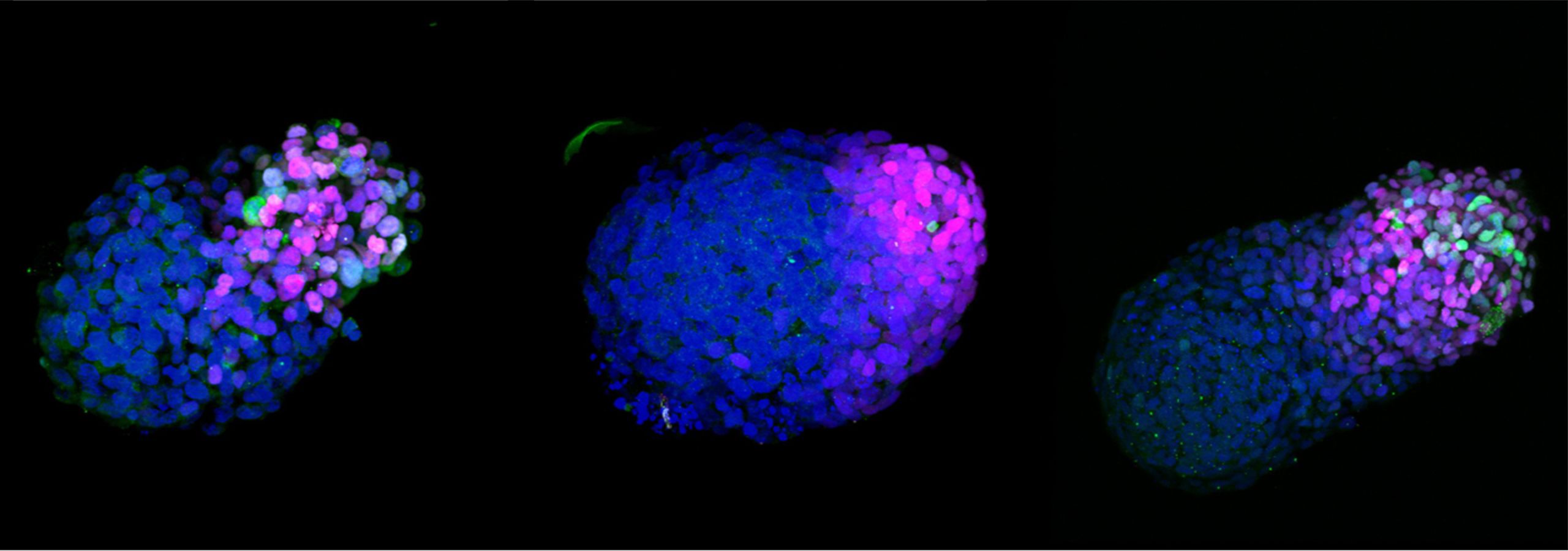
A growing human gastruloid at 24 hours (left), 48 hours (middle) and 72 hours (right). Blue marks DNA, magenta marks neural cells, green marks mesodermal cells.
A growing human gastruloid at 24 hours (left), 48 hours (middle) and 72 hours (right). Blue marks DNA, magenta marks neural cells, green marks mesodermal cells.

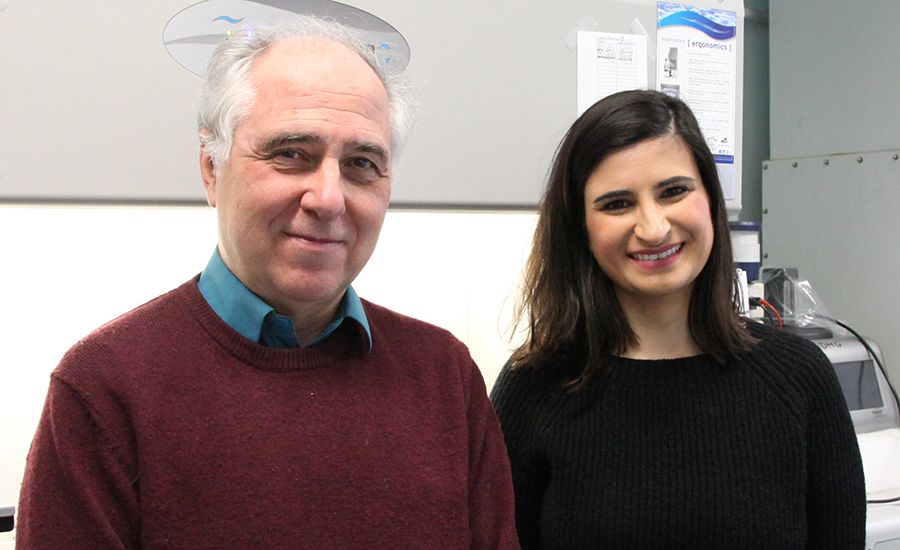
Professor Alfonso Martinez-Arias and Dr Naomi Moris, who led the research, in the Department of Genetics at the University of Cambridge
Professor Alfonso Martinez-Arias and Dr Naomi Moris, who led the research, in the Department of Genetics at the University of Cambridge
Human embryos as the source of human embryonic stem cells
Real embryos are formed when an egg and sperm fuse, in the process called fertilisation. Embryo-like structures are created in the lab from embryonic stem cells – cells from a human embryo at three to five days old. The human embryos are donated for research with consent; some of these come from eggs that were fertilised at in vitro fertilisation clinics but not used. The embryonic stem cells extracted from these embryos have the ability to develop into any type of specialised cell in the body, or can divide into more stem cells.
The 14-day rule
Researchers in this field follow an internationally acknowledged ethical guideline called the 14-day rule, first recommended in 1984. This limits research on human embryos to the two-week period after fertilisation. The limit relates to the perceived moral status of the human embryo, judged to be acquired during the process of development. In addition, 14 days is roughly the time that gastrulation occurs in the embryo.
Applying the 14-day rule to research on embryo-like models
In the last few years, various new techniques have been developed to produce embryo-like models from animal and human stem cells. This has driven researchers to call for specific guidelines to provide clearer ethical oversight of this rapidly developing field.
Different countries have different approaches to research on embryo-like models derived from human stem cells. In the US, Federal research is restricted in the same way as for human embryos (US Federal law bans the US government from funding research that creates or destroys human embryos). In other countries including the UK and Japan, research on embryo-like models is allowed, because they do not have the potential to grow into an individual.
This research was funded by the Medical Research Council, the Leverhulme Trust and the Isaac Newton Trust. It was reviewed and approved by the Ethics Committee of the University of Cambridge.
Reference Moris, N. et al. ‘An in vitro model for anteroposterior organisation during human development.' Nature, June 2020. DOI:10.1038/s41586-020-2383-9
Image credits Naomi Moris, University of Cambridge. Black and white SEM image made with the assistance of K. Muller at the Cambridge Advanced Imaging Centre.
Media enquiries Jacqueline Garget
Researcher contacts Professor Alfonso Martinez-Arias, Dr Naomi Moris
