A shake of the dice and a nod from the neighbour – new facets of stem cell biology uncovered when methods in theoretical physics were used to solve a biological problem.
A shake of the dice and a nod from the neighbour – new facets of stem cell biology uncovered when methods in theoretical physics were used to solve a biological problem.
We depend on the ability of our cells to renew and repair, both for when we are injured and for the normal maintenance of tissues like skin that suffer continual cell loss.
Physicist Professor Ben Simons, newly appointed Herchel Smith Professor of Physics of Medicine, has spent the past two decades working with ultracold atomic gases and quantum chaos. But his research took a biomedical turn after a chance conversation with clinician and cancer researcher Dr Phil Jones, the results of which have shaken up long-held views about how stem cells behave and how tissues renew themselves.
A perfect balance
We depend on the ability of our cells to renew and repair, both for when we are injured and for the normal maintenance of tissues like skin that suffer continual cell loss. This astonishing capacity for regeneration is down to our stem cells, which are able both to renew themselves indefinitely and also to give rise to daughter cells that mature into the multitude of different cell types needed to replenish tissue.
‘Key to this process is the perfect balance that tissue stem cells must maintain between cell loss and cell replacement. Working out the mechanism that controls the balance represents one of the defining questions of tissue stem cell biology, not least because too much cell production could cause cancer, and too little could contribute to tissue failure,’ explains Professor Simons.
The prevailing view has been that the balance is maintained because a long-lived slow-cycling stem cell divides asymmetrically to produce a copy of itself and a daughter cell that matures – at once maintaining the stem cell population and replacing the cell lost from the tissue. However, using an innovative lineage tracing technique and concepts from statistical physics, Professor Simons and his collaborators have amassed data that suggest a new paradigm for stem cell behaviour.
Throwing dice
In 2001, Dr Phil Jones at the Medical Research Council (MRC) Cancer Cell Unit had set out to confirm the theory that stem cells divide in an asymmetrical fashion so that he could use it as a platform to study mechanisms of dysregulation and cancer progression.
‘Normal cells follow a remarkably simple set of mathematical rules of behaviour that become distorted in cancer,’ he explains. ‘Once we know what the rules are, we can test how drugs alter cell behaviour and apply this to understanding how to prevent cancers developing and improve cancer treatment.’
He used a remarkable tool for tracking what stem cells do: a transgenic mouse made by Dr Douglas Winton at the Cancer Research UK Cambridge Research Institute that carries a genetic reporter which can be induced to fluoresce. Using 3D imaging, it is possible to trace what happens to the lineage that individual stem cells create.
After several years of observations, the result was an enormously complex dataset. It was at this stage that Simons became involved. As a physicist, he realised the dataset had a certain familiarity: ‘For all its complexity, there was a simple feature of the dataset that physicists would recognise as scaling. Once we spotted this, we knew that the data didn’t fit the predictions of the paradigm.’
‘Most cells expand by doubling, producing 2, 4, 8 daughter cells and so on,’ adds Jones. ‘But we found that these cells followed a 1, 2, 3, 4 growth model, where on average one of the daughter cells stops growing while the other carries on.’
It seems that the cells are not dividing in the asymmetrical fashion expected of stem cells: instead, they produce two identical copies of themselves, or two identical cells that mature, or one of each. ‘These cells are behaving rather like they are throwing dice,’ Simons explains. ‘Of course this dice throwing game is the product of very complex regulation but the outcome is both unpredictable and perfectly balanced between three possible fates.’
What the neighbours do
Inspired by the success of the collaboration with Jones, the results of which were published in Nature, Simons began to look through the scientific literature for examples of similar datasets – very high-quality lineage tracing experiments yielding vast amounts of data – and discovered the work of Dr Shosei Yoshida from the National Institute for Basic Biology in Okazaki, Japan.
Dr Yoshida had been looking at another instance of a tissue that is constantly renewed – the mammalian spermatogenesis system. With the help of an MRC Discipline Hopping grant, Simons embarked on a statistical analysis in collaboration with Yoshida and found evidence for the same unpredictable fate behaviour similar to that seen in skin. This time, however, stem cells symmetrically self-renew in response to the chance loss of a neighbour, leading to a random drift of the stem cell population.
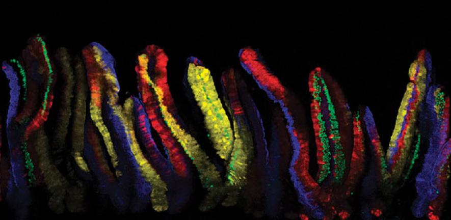
Encouraged by these findings, Simons and Winton turned to the fastest self-renewing tissue in mammals – the lining of the intestine. Here, the burden of replenishing the millions of cells that are lost daily falls on a small group of stem cells at the base of the intestinal crypts.
Once again, by tracing the lineage of dividing stem cells, they were able to define the pattern of stem cell fate. ‘It’s amazingly simple,’ Simons explains. ‘In effect, all cells have the same ‘life chances’; if the roll of the dice determines that it will differentiate and move out of the crypt then it is replaced by a stem cell that simply divides into two stem cells.’ Following collaboration with Professor Hans Clevers, from the Hubrecht Institute in Utrecht, vivid evidence of stem cell dynamics has now been found using a multicolour labelling system in mice.
Physics, stem cells and cancer
Following that serendipitous conversation five years ago, Simons has built a host of collaborative activities that use methods in theoretical physics to solve biological problems. Fittingly, his professorship is associated with the Physics of Medicine initiative (www.pom.cam.ac.uk/), which aims to establish and co-ordinate new interdisciplinary activities between the physical and life sciences, and he now contributes to both Cambridge’s Stem Cell Initiative (www.stemcells.cam.ac.uk/) and the thriving cancer research community (www.cambridgecancercentre.org.uk/).
His ongoing collaborations with stem cell and developmental biologists are now looking at other adult tissues and also at carcinogenesis. So far, all tissues Simons and his collaborators have looked at show the same characteristic stem cell behaviour.
‘This tells us important information about the rules of normal stem cell biology,’ says Simons. ‘We can for instance use this as the basis to interrogate the action of drugs and to understand what goes wrong in cancers. Cancer cells clearly have a growth advantage, but what is the origin of that advantage?
There is a classical view about the progression of tumours that postulates a sequence of mutation events – we are now in a position to address at single cell resolution what is really going on.’
For more information, please contact Professor Ben Simons (bds10@cam.ac.uk) who currently works between the Cavendish Laboratory of the Department of Physics and the Wellcome Trust and Cancer Research UK (CRUK) Gurdon Institute, and also has an adjunct faculty position at CRUK Cambridge Research Institute.

This work is licensed under a Creative Commons Licence. If you use this content on your site please link back to this page.

. Trophoblast cells are invading out of the organoid, mimicking placental cells invading the uterus in the early weeks of pregnancy., Credit: Friedrich Miescher Institute/University of Cambridge")

")