Medical imaging in Cambridge is pushing the boundaries in diagnosis and therapy as well as helping scientists within their own disciplines.
Medical imaging in Cambridge is pushing the boundaries in diagnosis and therapy as well as helping scientists within their own disciplines.
Today, imaging is a crucial part of the biomedical sciences and a mainstay of medical practice. No longer a scarce resource, imaging is often the first port of call for diagnosis, and now increasingly for treatment.
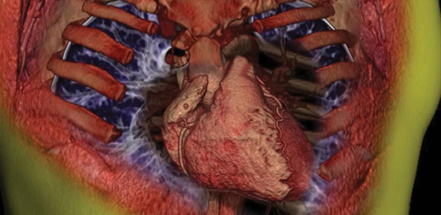
Medical imaging has developed at an astonishingly rapid speed since the first discovery of X-rays in 1895. Today, imaging is a crucial part of the biomedical sciences and a mainstay of medical practice. No longer a scarce resource, imaging is often the first port of call for diagnosis, and now increasingly for treatment. The modern-day repertoire of imaging techniques has widened from X-rays and now embraces ultrasound (US), computed tomography (CT), magnetic resonance imaging and spectroscopy (MRI/MRS), and nuclear medicine (NM; a term that includes positron emission tomography, PET).
In Cambridge, a close cooperation between research and medicine sees experienced researchers with technique and body system expertise, many of national and international acclaim, collaborating directly with specialty-based clinicians. A principal aim of much of this research is to translate novel imaging-based diagnostics and treatments from the laboratory into clinical practice, and to use imaging methods to monitor their effects.
Imaging for diagnosis
Medical imaging is nowadays relatively non-invasive for the patient and also speeds up their medical care. In the Department of Radiology, imaging techniques are being refined for diagnosing abdominal pain, cranial and spinal problems, early lung cancer, inflammatory bowel disease and arthritis, to name but a few conditions that are benefitting from these non-invasive diagnostic procedures. Image-guided strategies are replacing formal open surgical diagnostic procedures for many diseases. And, where direct injection of contrast material was previously required to visualise the lymphatic system, lymph nodes can now be directly visualised in three-dimensional images on ultrasound, CT, PET/CT and MR.
Imaging for therapy
As well as breaking new ground in how medical conditions are diagnosed, ongoing research is enabling the replacement of some forms of therapeutic surgery. This has been achieved in several conditions through the development of novel image-guided techniques that allow drainage of acute abdominal and pelvic abscesses. In breast malignancy, sophisticated percutaneous image-guided methods have been extended to incorporate therapeutic excision using wide-core needle biopsy, and this may, in time, replace conventional methods of breast surgery.
Imaging can inform surgeons, helping them to decide whether to operate, as well as enlighten the patient and guide surgical planning. These improvements to therapeutic surgery have been made possible through triage studies on acute surgical admissions using rapid multi-detector CT, providing surgeons with a pre-operative specific diagnosis and location of disease.
Towards novel imaging techniques
New and innovative uses of imaging are being pioneered and evaluated in Cambridge for a plethora of disease conditions. In patients with atherosclerosis, lesions in carotid arteries are being assessed using a variety of techniques that can also determine the degree of associated inflammatory change. Important nerve tracts are being delineated in relation to brain tumours, potentially improving tumour resection and radiotherapy outcomes and predicting likely impairments following surgery; and neuroradiologists are developing rapid techniques for evaluating patients with stroke. New functional methods such as MR perfusion and diffusion are being evaluated for assessing early solid tumour response to therapy (e.g. in gynaecological tumours). For diffuse liver disease, a range of quantitative techniques for measuring body and hepatic fat distribution are also being assessed.
Expanding horizons
Imaging is very much at the forefront of modern medicine, and any successful biomedical campus today is underpinned by first-class radiology and pathology. Despite the immense breakthroughs afforded by medical imaging, we seem to be nowhere near exhausting the vast potential of these techniques. For all stages of patient care, from screening to diagnosis, delivering therapy to monitoring outcome, the medical horizons for imaging continue to expand.
For more information, please contact the author Professor Adrian Dixon (akd15@radiol.cam.ac.uk) at the Department of Radiology.
Medical imaging across Cambridge
Cambridge is fortunate in having excellent cross-sectional imaging facilities that benefit from a close integration between National Health Service (NHS) departments and academic imaging departments spread across the city.
In the University’s Department of Radiology, located at Cambridge University Hospitals NHS Foundation Trust, Addenbrooke’s Hospital, an impressive inventory of imaging tools has been assembled thanks to research collaborations both with industry (GlaxoSmithKline) and with charity (Cancer Research UK; CRUK). The Department of Radiology houses numerous modern full-capability US systems, along with three multi-detector CT systems, four state-of-the-art 1.5 Tesla (T) MRI systems and one 3T MRI system. Two of the MRI systems have full multi-nuclear spectroscopy capability. These NHS-based machines are all used extensively for research as well as for routine NHS work. All machines are linked by a sophisticated network to a central picture-archiving computed storage (PACS) system, allowing practitioners at different physical locations to access the stored images.
Also used extensively for research as well as for routine NHS work are the gamma cameras within the NHS Department of Nuclear Medicine; the imminent arrival of a PET/CT unit in this Department, funded with a grant from the recent successful National Institute for Health Research (NIHR) Biomedical Research Centre bid and industrial support, will be predominantly used for research. Comprehensive research agreements with the vendors underpin the ability to develop and evaluate new technology and applications on these systems.
The Wolfson Brain Imaging Centre (WBIC) is a research facility attached directly to the Addenbrooke’s Hospital Neuro Critical Care Unit and is dedicated to imaging function in the injured human brain. WBIC facilities include state-of-the-art MRI/MRS systems (two of them 3T systems), PET research capabilities, two cyclotrons and a PET radiochemistry laboratory with some 15 radioligands currently available for studying patients with acute brain injury.
The University is currently refurbishing a laboratory on the Cambridge Biomedical Campus that will provide the School of Clinical Medicine with small animal imaging systems to support phenotyping and molecular imaging studies for metabolic, endocrine, neuroscience and cardiovascular medical research. In addition to a 4.7T preclinical MR and microPET, it will also house an experimental PET/MR system.
The Medical Imaging Group in the Department of Engineering is developing new techniques for freehand three-dimensional ultrasonic imaging. Working closely with various members of the School of Clinical Medicine, the Group is researching ways to improve the visualisation and measurement of tumours and more accurate targeting of the tumour site, allowing reduced radiation doses with fewer side effects.
The Cambridge Research Institute (CRI), funded by CRUK, has recently developed an Imaging Section and there is close liaison between its imaging scientists and those elsewhere on the campus. Imaging goals at the CRI include using MRI and MRS for the evaluation and design of novel tumour therapies, including immunotherapy, anti-vascular and gene therapies. Such research will increase understanding of the biology of cancer and the determination of tumour-associated MR parameters for diagnosis, prognosis and monitoring of therapy.
There are further imaging facilities at the Medical Research Council (MRC)-funded Cognition and Brain Sciences Unit, an internationally leading centre for research in the cognitive sciences and neurosciences. With dedicated 3T functional MRI and 306-channel magnetoencephalography facilities available on site, the Unit has particular strengths in the application of neuro-imaging techniques in the context of well-developed neuro-cognitive theory.

This work is licensed under a Creative Commons Licence. If you use this content on your site please link back to this page.


