Recent advances in medical imaging are being applied to airborne remote sensing of vegetation, enabling conservation scientists to see the wood and the trees.
Recent advances in medical imaging are being applied to airborne remote sensing of vegetation, enabling conservation scientists to see the wood and the trees.
It’s finally approaching what conservation scientists need to have: seeing the wood and the trees
David Coomes
Soaring over the tree canopy of one of the most biodiverse forests on earth, a tiny unmanned plane buzzes quietly through the air. Its pilot stands 250 m below, controlling its flight remotely. This unmanned aerial vehicle (UAV) is gathering data essential to understanding and diagnosing the health of the rainforest below.
The plane is one of a small fleet currently undergoing test flights in Indonesia. Each has been equipped with remote sensors. Their task is to image both the Harapan Rainforest – a 100,000 hectare area of formerly logged forest that is now managed for conservation by a group of NGOs including the RSPB – and a highly threatened forested area on the coast of Kenya.
Globally, around one billion hectares of degraded tropical forest like Harapan might be restorable, enabling them to continue to contribute to the planet’s biodiversity and its carbon and water cycles. But a major problem faced by conservation managers is how to survey extensive areas in which conditions can vary in just a few hundred square metres and are continually changing through natural regeneration.
A group of conservation scientists at the Department of Plant Sciences, RSPB and A Rocha International (which works in Kenya) has embarked on what it hopes is a cost-effective and high-quality solution, funded by the Cambridge Conservation Initiative Collaborative Fund. Lead researcher Dr David Coomes, explained: “Forest conservation activities often rely on airborne monitoring and satellite imagery to provide information but these are either expensive or don’t offer a fine-enough resolution. We’ve decided to use inexpensive sensors on UAVs to spot areas of the trashed forest that are showing early signs of recovery.”
The researchers need to measure the health of the forest on a tree-by-tree basis – locating, identifying and counting key species indicative of recovery. Multiply this up by hundreds of thousands of hectares, repeated at time intervals in the future, and it becomes a huge imaging, computational and ‘big data’ challenge.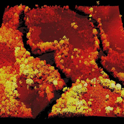
As the datasets grow, being able to manage and analyse the images automatically and with very high accuracy becomes crucial, and so a key part of the project is to develop the mathematical tools that will do this. This is the job of Dr Carola Schӧnlieb and her team of digital image analysts at the Department of Applied Mathematics and Theoretical Physics.
The mathematical tools have similarities to technology they are also developing for tumours. For the past year, Schӧnlieb’s group has been working on the VoxTox Research Programme – a five-year study funded by Cancer Research UK and led by Professor Neil Burnet at Cambridge’s Department of Oncology – which aims to reduce the ‘collateral damage’ toxicity that can arise during cancer radiotherapy.
About half of all people with cancer receive a course of radiotherapy, a form of treatment in which X-rays are used to shrink or destroy the tumour. With the benefit of advanced systems, it’s now possible to aim radiation beams at tumours more effectively than ever before, allowing increasing doses of radiotherapy with increased cancer cure rates, and also reducing side effects.
However, although clinicians use planning software to define the target area for treatment and deliver the optimal dose, any dose that falls outside the target area – for instance due to the positioning of the patient and their internal organs during treatment – can cause permanent and severe damage to normal tissues. VoxTox, which brings together cancer specialists, mathematicians, radiologists, physicists and engineers, is developing a set of tools that can be used to provide patients with the optimal dosage for their condition.
“The similarity between what we are doing in VoxTox and forest mapping is the development of mathematical algorithms that combine datasets – a process called registration – and then segment them into objects of interest,” explained Schӧnlieb.
For VoxTox, imaging data gathered during the course of a patient’s radiotherapy is analysed mathematically pixel by pixel (or, in fact, ‘voxel by voxel’ because it’s in three dimensions) within the patient outline, and the dose is then re-computed at that point, each day, during treatment.
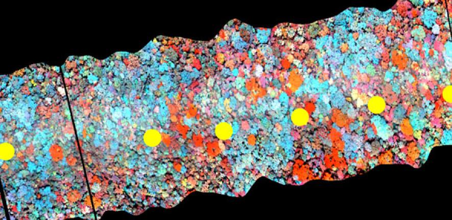
Airborne remote sensors for conservation, by contrast, gather data on which trees are in the forest, where they are and how healthy. “It’s like an airborne well tree clinic,” said Coomes. The data might include digital photography to record what can be seen; a three-dimensional laser scanner (or LiDAR) to measure the height of the canopy; and hyperspectral scanners to monitor the wavelengths of radiation each plant absorbs – these ‘chemical signatures’ can be used to identify species.
“Being able to bring these datasets together gives us a much fuller idea of the health of the forest than each of the datasets individually,” he added. “With the addition of GPS too, it means we can map the forest tree by tree, over time, in three dimensions.”
To develop the algorithms, researchers led by Schӧnlieb and Coomes are using test data previously acquired by Coomes’ group using manned flights over five European sites, as well as data recently gathered from 200 km2 of Malaysian forest as part of a project funded by the Natural Environment Research Council.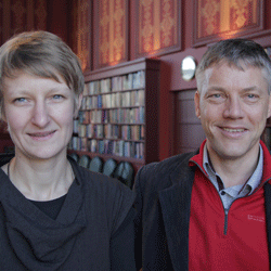
This is complex mathematical image processing, as Schӧnlieb explained: “As for VoxTox, the aim is to faultlessly match different types of sensing data as a hybrid dataset and then segment it based on the different levels of information present in each voxel. This sort of analysis hasn’t been done before with this kind of accuracy. It pushes over the boundaries of state-of-the-art methods.
“The next stage is to see how far we can push the segmentation method, which is the part that identifies individual trees,” she said. “If we can maintain high levels of accuracy using cheaper and fewer sensors – like those being used on the UAVs – then you can take imagery that’s as good as it’s going to get and maximise the Meanwhile information gain from what you have.”
Meanwhile, by mid-2015, the UAVs will begin streaming data back to the researchers who, with their algorithms ready, will start mapping the voxel forest and feeding the results into its management. “The appeal of this technology is you are dealing with individual plants and trees,” said Coomes, “it’s finally approaching what conservation scientists need to have: seeing the wood and the trees.”
Inset image – top: LiDAR image from Sierra Leone showing the Moru river along the border between Sierra Leone and Liberia; Credit: RSPB
Inset image – bottom: Dr Carola Schönlieb and Dr David Coomes; Credit: University of Cambridge
The text in this work is licensed under a Creative Commons Licence. If you use this content on your site please link back to this page. For image rights, please see the credits associated with each individual image.


