Teenage years can be difficult enough, but for people affected by schizophrenia, this is when symptoms first tend to arise. Dr Kirstie Whitaker (Department of Psychiatry), who is speaking at this year's Cambridge Science Festival, explains in The Conversation how her work may shed light on why this is the case.
Teenage years can be difficult enough, but for people affected by schizophrenia, this is when symptoms first tend to arise. Dr Kirstie Whitaker (Department of Psychiatry), who is speaking at this year's Cambridge Science Festival, explains in The Conversation how her work may shed light on why this is the case.
When I was studying for my PhD at the University of California at Berkeley, I spent an awful lot of my weekends asking teenagers to lie still in a magnetic resonance imaging (MRI) scanner. While they were lying as still as they could they also had to answer questions they saw on the screen. ![]()
We were interested in which parts of the brain these young people were using when they completed an analogical reasoning task. They saw three pictures and were asked to choose the fourth picture to complete the relationships, for example dress is to wardrobe, as milk is to fridge.
We found that association cortex – the parts of the brain that bring together information from many other regions – was used to answer these questions and, importantly, that these regions are activated more and more as you get older. The study was recently published in Developmental Science.

Fast forward a few years and I’m now a member of the Neuroscience in Psychiatry Network (NSPN), a collaboration between the University of Cambridge and University College London, funded by the Wellcome Trust.
NSPN aims to better understand the biological mechanisms that lead to young people developing a variety of mental health disorders. The network includes experts in epidemiology, adolescent psychopathology, cognition and brain development who are investigating the question of adolescent mental health from multiple angles.
Beautiful brains
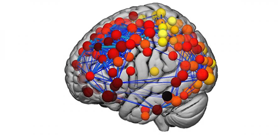
I’m in the MRI team, which has collected brain scans from 300 young people between the ages of 14 and 24. This time, instead of asking them to complete questions while they were lying in the scanner, we took structural MRI scans. These are different to the brain scans we use to assess what the brain is doing (“functional MRI”) and they look really beautiful.

One of the measures we extract is “cortical thickness” – the depth of the outside layer of the brain (the cortex) that contains synapses. A synapse is where two brain cells (neurons) join together and transmit messages. We have around 100 billion neurons but 100 trillion synapses in our brains. It is the complexity of these connections that allows humans to generate and understand complex thoughts and feelings, including being able to solve analogies in the real world.
Changing minds
In work published in the Proceedings of the National Academy of Science in 2016 the NSPN consortium showed that the cortex got thinner between the ages of 14 and 24. In particular, association cortex – the same regions that are used for complex thought and reasoning – showed the greatest amount of change.
This finding may seem counter-intuitive – at first glance you’d imagine that getting better at something would mean having more brain cells working on the project. In fact, you are born with more neurons than you’ll ever have again, and one of the most important developmental processes is “synaptic refinement” – pruning away some of the connections to ensure your brain is working efficiently.
We also looked at the brain as a network. You can imagine an airline network with connections going around the world between different airports. The really important locations are network “hubs” – they have lots of flights in and out every day. In the brain, we found that these hubs are located in association cortex. It makes sense that the brain regions that are important for complex thought need information to liaise with many other different parts of the brain.
We found that the hubs of the brain’s structural network change most during adolescence. This is likely to reflect prolonged development for these regions – the other parts of the brain are closer to their adult structure and have slowed down by the age of 14. If you think back to my descriptions of synapses being pruned away, it makes sense to keep as many as you need for a long time, until you’re sure of which connections are going to allow you to best reason in the complex world around you.
I made a big deal earlier about the fact that we can not see the actual cells and connections in the brain using MRI. However, innovative work by Petra Vértes and data shared by the Allen Institute for Brain Science gives us some clues as to what is going on at the cellular level in these brain regions.
The Allen Institute has measured the expression of 20,000 genes at 500 locations around the brains of six brains donated to research after their owner died. Vértes showed that the brain regions in association cortex – the same ones that change structurally the most during the teenage years – have greater expression of genes related to synaptic plasticity. This means that genes controlling how our brain cells adapt their connections based on our experiences are more active in these regions.
These regions are also related to genes associated with schizophrenia – a psychiatric disorder that is most likely to emerge during the late teenage years. Our work provides evidence for a mechanism as to why people with a genetic risk for schizophrenia may not experience symptoms until they are around 20-years-old. The affected brain regions are still developing and it may take these many years before the differences in brain structure are big enough to cause the hallucinations and delusions associated with this mental health disorder.
There is still a long way to go in our search to understand the biological underpinnings of mental health disorders. We will continue to work together, both within the Neuroscience in Psychiatry Network and with other researchers around the world, to find treatments for mental health disorders, and, if possible, to find ways to prevent the symptoms from emerging all together.
The author is appearing on March 21 as part of the Cambridge Science Festival.
Kirstie Whitaker, Research Associate, Department of Psychiatry, University of Cambridge
This article was originally published on The Conversation. Read the original article.

The text in this work is licensed under a Creative Commons Attribution 4.0 International License. For image use please see separate credits above.

")

